

1 Introduction: A Major Application Area for Potentiostats in Biosensor Research
When researchers discuss potentiostats, the conversation tends to gravitate towards batteries, green hydrogen, and corrosion — disciplines with large capital investment, national policy backing, and obvious industrial scale. Biosensing rarely leads that conversation. Yet by market share, it dominates.
Market-report estimates suggest that biosensing is a major application segment for wireless potentiostats, with some reports placing it at approximately 35% of market revenue. Personal glucose meters used by hundreds of millions of people with diabetes worldwide are a familiar example of electrochemical biosensing. Research-stage biosensors also use amperometric, voltammetric, potentiometric, and impedance-based readouts for biomarkers, pathogens, and nucleic-acid targets.
Every one of these devices, at some point in its development, was characterised, validated, and optimised using a potentiostat.
This guide covers the complete electrochemical workflow for biosensor research: how DPV, SWV, and EIS work as analytical techniques, how to select the right electrode platform for your application, and what the transition from a laboratory potentiostat experiment to a deployable diagnostic device actually involves in practice — particularly for AU/NZ pharmaceutical, clinical diagnostics, and environmental health research groups.
If you are new to the physical setup of an electrochemical experiment — including electrode preparation, lead connections, and OCP monitoring — the starting point is our potentiostat setup guide for 3-electrode cells. For a broader view of how potentiostat electrochemistry operates across AU/NZ research sectors, see our sector overview guide.
2 Why Electrochemical Detection Works So Well for Biosensors
Biosensor detection platforms are broadly classified by their transduction mechanism — the physical process by which a molecular recognition event is converted into a measurable signal. Optical biosensors use fluorescence or absorbance. Mass-based sensors use piezoelectric resonance. Electrochemical biosensors use electrical current, potential, or impedance.
Electrochemical transduction has dominated the commercial biosensor market for four decades for reasons that are straightforward when you think about them from a deployment perspective:
- Miniaturisation — The electronic circuitry required for electrochemical measurement can be miniaturised onto a chip. Depending on the design and application, modern potentiostat-on-a-chip platforms can be fabricated into compact devices with wireless communication, sensitivity suitable for selected clinical analyte ranges, and low-power operation. Optical and mass-based transduction systems face substantially harder engineering challenges at equivalent scale.
- Sample matrix compatibility — Electrochemical techniques work directly in complex biological matrices — blood, urine, saliva, sweat — without the sample clarification steps that optical methods often require. The working electrode surface is what matters; the optical properties of the sample do not interfere.
- Quantitative precision — The current or impedance response of an electrochemical biosensor is directly related to analyte concentration through well-defined physical laws. This makes calibration straightforward and the measurement inherently quantitative rather than qualitative.
- Cost of consumables — A thick film screen-printed electrode incorporating the full three-electrode configuration costs a fraction of the equivalent optical transducer. Disposability eliminates the fouling, regeneration, and cross-contamination challenges that reusable sensor platforms must manage.
These four properties — miniaturisation, matrix compatibility, quantitative precision, and consumable economics — are why electrochemical biosensors command their market position. They are also why the potentiostat is the central instrument in any biosensor development programme.
People Also Ask: Why Are Electrochemical Biosensors More Common Than Optical Biosensors?
Electrochemical biosensors dominate the commercial market because their transduction circuitry miniaturises readily onto chips, they work directly in complex biological matrices without sample clarification, they produce inherently quantitative signals calibrated against well-defined physical laws, and their disposable electrode consumables (screen-printed electrodes) are significantly cheaper than equivalent optical transducers. These properties make electrochemical detection the preferred platform for point-of-care diagnostics, wearable health monitors, and field-deployable environmental sensors.
3 The Three Core Techniques: DPV, SWV and EIS
The potentiostat enables a repertoire of electrochemical techniques. For biosensor development and pharmaceutical analysis in the AU/NZ context, three techniques account for the vast majority of published research and applied diagnostic work. Understanding what each one measures — and where each one’s advantage lies — is essential before choosing which to build your assay around. For a broader overview of all potentiostat techniques across research sectors, see our potentiostat applications guide.
Differential Pulse Voltammetry (DPV): The Sensitivity Benchmark
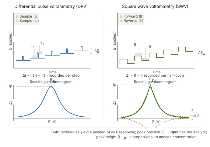
Differential Pulse Voltammetry (DPV) is the workhorse technique for trace analyte detection in complex biological and pharmaceutical samples. The potentiostat applies a series of small potential pulses superimposed on a linearly ramping baseline voltage, and measures the difference in current immediately before and immediately after each pulse.
This differential measurement is the technique’s defining advantage. By measuring the change in current rather than its absolute value, DPV effectively cancels out the slowly varying capacitive background current — the dominant noise source in voltammetric measurements in complex matrices. The result can be a significant improvement in signal-to-noise ratio compared with simple linear sweep or cyclic voltammetry, enabling very low detection limits — including picomolar and, in some well-optimised systems, femtomolar ranges — depending on electrode design, surface chemistry, matrix, and assay protocol.
Where DPV is the right choice:
- Detection of electroactive biomarkers (dopamine, uric acid, ascorbic acid, adenine, guanine) in biological fluids
- Pharmaceutical impurity profiling — detecting electroactive degradation products or synthesis byproducts in active pharmaceutical ingredients at ICH Q3A/Q3B concentration thresholds
- Nucleic acid hybridisation detection — labelled DNA or RNA targets produce a DPV peak at a characteristic potential on a functionalised screen-printed electrode
- Heavy metal stripping analysis in environmental samples — anodic stripping DPV of lead, cadmium, and arsenic in drinking water using 3D porous graphene sensing strip electrodes
Key parameters to optimise in DPV: Pulse amplitude (typically 25–50 mV), pulse width (10–100 ms), step potential (5–10 mV), and scan rate. The combination of pulse amplitude and step potential determines the effective scan rate and the balance between sensitivity and peak resolution. For pharmaceutical work, lower step potentials (5 mV) give better resolution of closely spaced oxidation peaks from structurally similar impurities.
Square Wave Voltammetry (SWV): Speed Without Sacrificing Sensitivity
Square Wave Voltammetry (SWV) applies a symmetric square-wave oscillation on top of a staircase potential ramp, and records the difference between the forward (anodic or cathodic) and reverse current components at each step. Like DPV, the differential measurement rejects capacitive background — but SWV achieves this at substantially faster scan rates.
Where DPV may require tens of seconds to several minutes per scan, SWV can often complete an equivalent potential range more rapidly, depending on the frequency, step size, potential window, electrode platform, and instrument settings. This speed advantage becomes critical in two contexts:
- Real-time monitoring applications — Continuous amperometric glucose monitoring, lactate tracking in elite sport science, and continuous environmental sensor networks all require measurement cycles short enough not to miss transient concentration spikes. SWV provides the sensitivity of DPV with the throughput needed for these dynamic monitoring scenarios.
- Enzyme-labelled immunoassays — Lateral-flow immunoassay formats with electrochemical readout (electrochemical ELISA) use SWV to interrogate enzyme-labelled antibody-antigen complexes. The faster scan rate reduces the total assay time, which matters in point-of-care contexts where time-to-result drives clinical utility.
-
Where SWV is the right choice: - High-throughput pharmaceutical screening — multiple compound libraries analysed at low sample volume on thin-film microfabricated electrodes - Aptamer-based biosensors where conformational switching upon target binding produces a measurable change in peak current — the speed of SWV is essential when monitoring real-time binding kinetics - Food safety analysis — rapid detection of antibiotics, pesticide residues, and mycotoxins in food matrices where analytical throughput determines commercial viability
People Also Ask: What Is the Difference Between DPV and SWV in Biosensor Research?
DPV (Differential Pulse Voltammetry) applies potential pulses on a linear ramp and measures the differential current, achieving picomolar detection limits with moderate scan speeds (60–120 seconds per scan). SWV (Square Wave Voltammetry) uses a symmetric square-wave oscillation and can achieve comparable sensitivity at substantially faster scan rates in many biosensor systems, depending on the method parameters and electrode configuration. DPV is preferred for maximum sensitivity in trace biomarker detection and pharmaceutical impurity profiling. SWV is preferred when throughput matters — real-time monitoring, high-throughput screening, aptamer-based sensors, and rapid point-of-care immunoassay formats.
Electrochemical Impedance Spectroscopy (EIS): Listening Without Speaking
Electrochemical Impedance Spectroscopy (EIS) approaches analyte detection from a fundamentally different direction to DPV and SWV. Rather than driving a faradaic reaction and measuring the resulting current, EIS probes the electrical properties of the electrode-solution interface by applying a small-amplitude sinusoidal perturbation — often around 5–10 mV — across a defined frequency range, such as from high-frequency kHz ranges down to low-frequency mHz ranges, depending on the instrument and experiment.
In the biosensor context, the power of EIS lies in what happens to the electrode-solution interface when a target molecule binds:
When an antibody, aptamer, or molecularly imprinted polymer immobilised on the electrode surface captures its target analyte, the captured molecule physically blocks the electrode surface — increasing the charge-transfer resistance (Rct) by a measurable and concentration-dependent amount. This is label-free detection: no enzyme, no fluorophore, no electroactive reporter molecule is required. The binding event itself is the signal.
The Nyquist plot interpretation for biosensor EIS: A Nyquist plot of a well-functionalised biosensor electrode typically shows: - A high-frequency semicircle whose diameter represents R_ct — the kinetic barrier to electron transfer through the electrode-solution interface - Increasing R_ct after each successive analyte concentration increment, tracked in a calibration series - A low-frequency Warburg diffusion tail representing mass transport of the redox probe (typically ferri/ferrocyanide couple, [Fe(CN)₆]³⁻/⁴⁻) to the electrode surface
The shift in R_ct between the bare electrode, the functionalised electrode, and the analyte-exposed electrode is the quantitative biosensor signal. Some well-designed EIS biosensors can achieve low detection limits for protein and nucleic-acid targets without requiring electroactive labelling, which can be a practical advantage for selected complex clinical sample matrices.
Where EIS is the right choice:
- Antibody-antigen (immunosensor) detection — the large size of antibody-antigen complexes produces substantial Rct increases measurable at clinically relevant concentrations
- Aptamer-based sensors for proteins (thrombin, VEGF, PSA) and small molecules (adenosine, cocaine, antibiotics) particularly where label-free operation is a regulatory or practical requirement
- Whole-cell and virus detection — intact microbial cells or viral particles produce distinctive impedance signatures upon capture at electrode surfaces
- Continuous, non-destructive monitoring — EIS does not consume the analyte or irreversibly alter the sensor surface, allowing repeated measurements on the same electrode
People Also Ask: How Does EIS Work for Biosensor Detection?
In EIS-based biosensor detection, a small sinusoidal voltage (5–10 mV) is applied across a range of frequencies (100 kHz to 10 mHz), and the resulting impedance spectrum is recorded. When target molecules bind to recognition elements (antibodies, aptamers, or MIPs) immobilised on the electrode surface, they block the electrode-solution interface and increase the charge-transfer resistance (Rct). This increase, measured from the Nyquist plot semicircle diameter, is proportional to analyte concentration. EIS offers label-free, non-destructive detection with detection limits comparable to DPV for protein and nucleic acid biosensors.

4 Electrode Platforms: Matching the Sensor to the Application
The potentiostat technique determines what you measure. The electrode platform determines where and how you measure it. Selecting the wrong electrode platform is one of the most common reasons that a biosensor assay which works in the lab fails to translate to a deployable format.
ScienceGears’ disposable electrochemical sensors range covers three distinct platform categories, each with specific strengths and application contexts.
Thick Film Screen-Printed Electrodes (SPEs) — The Universal Biosensor Platform
Thick film screen-printed electrodes are the most widely adopted electrode format in electrochemical biosensor research — and for good reason. ScienceGears distributes MicruX SPEs across Australia and New Zealand for biosensor, diagnostic, environmental, and electroanalytical research applications.
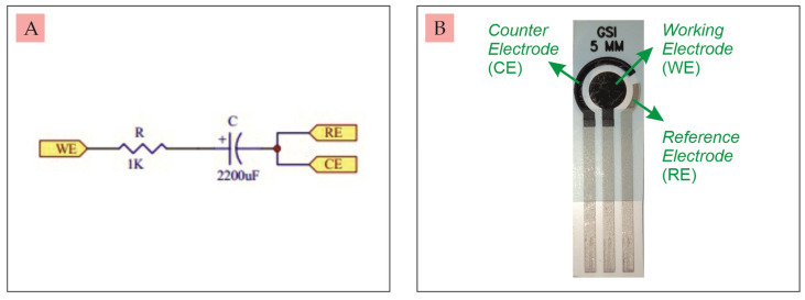
How they are made: Conductive inks — carbon, graphene, gold, or platinum composites — are precision-deposited onto rigid ceramic (Al₂O₃) or flexible PET polymer substrates using screen-printing, yielding a well-defined three-electrode configuration (working, reference, and auxiliary/counter electrode) in a single, ready-to-use, compact format.
Available working electrode materials and their biosensing applications:
- Carbon ink SPEs — The standard platform for most DPV and SWV analyte detection work. Carbon’s wide electrochemical window in aqueous media, low background current, and ease of surface modification (via electrodeposition, covalent chemistry, or drop-casting of nanoparticle composites) make carbon SPEs the default starting point for new biosensor development.
- Graphene ink SPEs — Enhanced electrochemical properties over carbon: higher electron transfer rate, larger electroactive surface area, and intrinsically lower oxidation overpotential for many biomarkers. Preferred for dopamine, uric acid, and ascorbic acid simultaneous detection applications where resolution of overlapping DPV peaks is critical.
- Gold SPEs — The platform of choice for thiol-based self-assembled monolayer (SAM) biosensor architectures. Gold-thiol chemistry is the most reproducible surface functionalisation strategy available for antibody and aptamer immobilisation — critical when regulatory reproducibility requirements apply. Preferred for EIS immunosensors and aptasensors.
- Platinum SPEs — Used where catalytic activity or hydrogen evolution/detection applications require platinum’s specific electrochemical properties. Less common in biosensing than carbon or gold but important for specific enzymatic electrode configurations.
3D Porous Graphene Sensing Strip Electrodes — High Surface Area for Trace Detection
3D porous graphene sensing strip electrodes represent the next generation of disposable electrochemical sensing platforms. Rather than a flat two-dimensional electrode surface, these strips incorporate a three-dimensional porous graphene architecture that increases the electrochemically active surface area available for analyte interaction and electron transfer.
The practical consequence is a significantly enhanced signal-to-noise ratio compared to conventional flat SPEs — making 3D graphene strips the preferred platform when trace-level detection is the priority:
- Environmental heavy metal analysis — Anodic stripping voltammetry for lead, cadmium, and arsenic detection at low-level concentrations in agricultural water, coastal marine sediment pore water, and drinking water, with achievable detection limits depending on electrode preparation, matrix effects, method validation, and instrument configuration
- Food safety — Simultaneous multi-metal determination in food matrices using a single DPV scan
- Pharmaceutical trace impurity analysis — Where ICH thresholds require detection at the low μg/g level in complex API matrices
Available across Australia and New Zealand through ScienceGears, with full technical support for application-specific electrode surface modification strategies.
Thin-Film Microfabricated Electrodes — Precision at the Microscale
Thin-film microfabricated electrodes are produced by lithographic deposition of electrode materials — gold, platinum, carbon — onto silicon or glass substrates at micrometre-scale dimensions. They represent the bridge between laboratory biosensor research and clinical diagnostic device fabrication.
Key advantages over thick-film SPEs:
- Dimensional precision — Lithographically defined electrode geometry can offer high dimensional precision and improved batch-to-batch consistency compared with many thick-film screen-printed formats, depending on the fabrication process and quality-control method.
- Integration with microfluidics — Thin-film electrodes are the natural electrode format for electrochemical and microfluidic platforms — lab-on-chip devices where sample volumes are in the nanolitre to microlitre range and channel dimensions are defined by photolithography.
- Array formats — Thin-film fabrication enables multi-electrode arrays on a single chip, enabling simultaneous multiplexed detection of multiple biomarkers from a single sample aliquot.
- Surface quality — The smoother, better-defined surface of a lithographically deposited gold electrode enables higher-quality SAM formation and more reproducible aptamer/antibody immobilisation than screen-printed surfaces.
For research groups moving from proof-of-concept DPV/SWV assay development on conventional SPEs towards a miniaturised, microfluidics-integrated diagnostic device format, thin-film microfabricated electrodes are the appropriate next step in the translation pathway.
5 The AU/NZ Biosensor Research and Diagnostic Development Landscape
Australia and New Zealand have active electrochemical biosensor research communities, with work spanning fundamental sensor science, applied diagnostics, environmental monitoring, and translational device development. Understanding where this work is happening, and what it needs from an instrumentation and consumable perspective, is what positions ScienceGears as a research partner rather than a product catalogue.
University Research Programmes
At QUT, the Centre for Materials Science has reported research on sustainable, biocompatible materials, including chitosan-derived wearable electronic transistor platforms from seafood waste, published in Small Structures in 2025. This type of work sits at the convergence of materials science, electrochemistry, biosensing, and wearable device development, and is relevant to researchers using electrochemical and spectroelectrochemical characterisation methods.
At UNSW, the Graduate School of Biomedical Engineering hosts active biosensor groups working on electrochemical detection of cancer biomarkers, sepsis markers, and infectious disease pathogens. The University of Melbourne’s Bio21 Institute and Monash Institute of Pharmaceutical Sciences both run programmes in electrochemical drug sensing and metabolite detection that depend on DPV and SWV as primary analytical techniques.
The Pharmaceutical Sector
Australia’s pharmaceutical and biotechnology sector — including established manufacturers, contract research organisations, and emerging biotech companies — is increasingly interested in electrochemical sensing for:
- Quality control of biopharmaceuticals — Electrochemical detection of oxidative modifications in monoclonal antibody formulations, where DPV identifies tryptophan and methionine oxidation products at concentrations relevant to stability specification limits
- Process analytical technology (PAT) — In-line electrochemical sensors for real-time monitoring of bioreactor redox state and metabolite concentrations during therapeutic protein production
- Drug-target interaction studies — EIS-based competition assays on aptamer-functionalised gold SPEs that quantify binding affinity constants for lead compounds in early-stage drug discovery
Point-of-Care Diagnostics and Remote Healthcare
Australia’s geography creates a specific and pressing need for field-deployable diagnostic tools. Remote communities across regional Australia and the Pacific Islands face infectious disease challenges — including elevated rates of tropical diseases, antibiotic-resistant infections, and nutritional deficiencies — where the current standard of care requires sample shipment to centralised laboratories with turnaround times measured in days.
A portable potentiostat paired with a functionalised screen-printed electrode can support rapid electrochemical immunoassay development, with assay time, sample type, and infrastructure requirements depending on the biorecognition chemistry, sample preparation, electrode design, and validation protocol. This is not a future technology — it is the direction that multiple AU/NZ diagnostic development programmes are actively pursuing, and the instrument and consumable combination that ScienceGears supplies is directly enabling that work.
People Also Ask: How Important Is Biosensing in the Potentiostat Market?
Market-report estimates suggest that biosensing is a major application area for wireless potentiostats, with some reports placing it at approximately 35% of market revenue.
| Sector | Primary Analyte Targets | Techniques | AU/NZ Institutions |
|---|---|---|---|
| Clinical diagnostics | Glucose, Troponin I, CRP, microRNA, SARS-CoV-2 antigen, Procalcitonin | DPV, EIS, Amperometry | Univ. of Queensland, Monash Univ., CSIRO Health & Biosecurity, ARC Centre CBNS |
| Pharmaceutical QC | Paracetamol, Ascorbic acid, Dopamine, Antibiotics, Uric acid | SWV, DPV, EIS | Univ. of Auckland, UNSW Sydney, Univ. of Melbourne, Metrohm ANZ |
| Environmental monitoring | Heavy metals (Pb, Cd, Hg), Nitrate/Phosphate, PFAS, Pesticides, Microplastics | SWASV, DPV, Amperometry | CSIRO Land & Water, GNS Science NZ, Curtin Univ., CRC CARE |
| Food safety | Salmonella, E. coli O157:H7, Aflatoxins, Histamine, Allergens | EIS, DPV, Amperometry | Food Science Australia, AgResearch NZ, Univ. of Adelaide, RMIT |
| Wearable / remote health | Sweat lactate, Cortisol, Urea, Na⁺/K⁺ electrolytes, Interstitial glucose | Amperometry, EIS, DPV | Univ. of Sydney, AUT Auckland, Griffith Univ., ARC ITTC FlexiSense |
Technique key: DPV = Differential pulse voltammetry · SWV = Square wave voltammetry · EIS = Electrochemical impedance spectroscopy · SWASV = Square wave anodic stripping voltammetry
6 Biosensor Development Workflow: From Assay Concept to Validated Sensor
The journey from an analyte detection idea to a validated electrochemical biosensor involves a sequence of discrete experimental stages — each of which has specific potentiostat technique and electrode platform requirements. Understanding this workflow helps in specifying the right instruments at the right stage of development rather than over-investing in system complexity before it is needed.
Stage 1 — Electrode Material and Surface Modification Screening
Use cyclic voltammetry on a conventional working electrode (glassy carbon or gold disc) to screen candidate surface modification chemistries — nanoparticle composites, conducting polymers, SAM architectures. Assess electroactive surface area by the Randles-Ševčík equation from the scan-rate-dependent peak current. This stage does not require disposable SPEs — conventional reusable electrodes on a benchtop single-channel potentiostat are appropriate.
Stage 2 — Bioreceptor Immobilisation and EIS Characterisation
Immobilise the recognition element (antibody, aptamer, enzyme, or MIP) onto the electrode surface. Use EIS to confirm each immobilisation step — the progressive increase in Rct as each layer is added provides direct, label-free evidence that the bioreceptor is present and properly oriented at the surface. Transition to MicruX gold SPEs for thiol-based SAM architectures at this stage.
Stage 3 — Analytical Performance Characterisation
Establish the calibration curve (signal vs concentration), limit of detection (LOD), limit of quantification (LOQ), linear dynamic range, and selectivity (against relevant interferents). Use DPV or SWV as the primary detection technique. For clinical applications, validate in spiked matrix (serum, urine, saliva) at relevant concentration ranges. 3D porous graphene strip electrodes offer enhanced LOD for analytes at sub-ppb concentrations.
Stage 4 — Integration with Portable or Microfluidic Platform
Transition from benchtop potentiostat to a portable potentiostat to assess performance under field-deployable conditions. Integrate SPEs or thin-film microfabricated electrodes into electrochemical and microfluidic platforms for sample handling integration. Assess performance with small sample volumes (1–50 μL) representative of finger-prick blood or swab eluate.
Stage 5 — Stability, Reproducibility, and Regulatory Data Package
Assess electrode-to-electrode reproducibility across production batches. Assess sensor storage stability (shelf life). Generate the analytical data package needed for TGA regulatory submissions or CE marking applications, depending on target markets.
The core potentiostat techniques in biosensor development are: (1) Cyclic voltammetry (CV) for electrode material characterisation and surface modification screening; (2) Electrochemical Impedance Spectroscopy (EIS) for label-free monitoring of bioreceptor immobilisation steps and analyte binding events; (3) Differential Pulse Voltammetry (DPV) for trace analyte detection with picomolar sensitivity; (4) Square Wave Voltammetry (SWV) for rapid detection with equivalent sensitivity to DPV at 5–10× faster scan rates; and (5) Chronoamperometry for real-time, continuous enzyme-based biosensor monitoring (glucose, lactate).

7 How ScienceGears Supports Biosensor and Diagnostic Research in AU/NZ
Biosensor development sits at an unusual intersection of analytical chemistry, materials science, biology, and engineering. The challenges that research groups encounter — inconsistent electrode functionalisation, irreproducible EIS spectra, and poor matrix performance despite clean standard-curve data — are rarely solved by reading the instrument manual alone. They are solved by talking to someone who has encountered the same problem in their own research and understands why it happens at the molecular level.
Dr. Siva Arumugam’s published research career has been built almost entirely at exactly this intersection. His work on SERS-based DNA hybridisation and mutation detection, electrochemical quantification of SERS enhancement factors, and related electrochemical sensing research reflects hands-on experience across electrochemical, spectroscopic, and biosensing workflows relevant to AU/NZ research groups.
When a biosensor group at a Queensland university contacts ScienceGears about setting up an EIS-based aptasensor for VEGF detection in serum, the conversation that follows is not a product recommendation drawn from a sales script. It is a scientific discussion about SAM quality on the gold SPE surface, about ferri/ferrocyanide probe concentration optimisation, about whether the R_ct shift they are seeing in PBS buffer will survive the matrix effects of diluted serum — and which potentiostat configuration will give them the noise floor they need to resolve the signal at clinically relevant concentrations.
That is what having co-founders with active research backgrounds in electrochemical sensing looks like in practice.
ScienceGears provides for biosensor research:
- Disposable electrochemical sensors — MicruX SPEs (carbon, graphene, gold, platinum), 3D porous graphene sensing strips, thin-film microfabricated electrodes
- Portable potentiostats for field-deployable and point-of-care validation
- Single-channel benchtop potentiostats for full DPV, SWV, EIS, and CV capability at the assay development stage
- Bipotentiostats for dual-electrode detection and RRDE measurements
- Electrochemical and microfluidic platforms and microfluidic accessories for lab-on-chip integration
- Spectroelectrochemistry cells for combined Raman/electrochemical characterisation of sensor surfaces
- Application notes, technical protocols, and direct consultation at sciencegears.com.au/application-notes
8 Pre-Experiment Checklist: Electrochemical Biosensor Assay Setup
Before running any DPV, SWV, or EIS biosensor measurement, verify the following:
- ☐ Electrode platform selected and appropriate for analyte, matrix, and target technique
- ☐ SPE or thin-film electrode stored correctly (humidity-controlled, dark) and within shelf life
- ☐ Buffer composition verified — pH, ionic strength, and redox probe concentration confirmed
- ☐ Electrode functionalisation steps completed and confirmed by EIS (Rct increase at each step)
- ☐ Potentiostat current range appropriate for expected peak currents (typically nA to μA for SPE biosensors)
- ☐ DPV/SWV parameters optimised on blank electrode in clean buffer before analyte introduction
- ☐ Blank measurement run completed to confirm no artefact peaks from buffer components or electrode impurities
- ☐ Calibration standard series prepared in appropriate matrix (buffer, spiked serum, spiked urine)
- ☐ Interferent selectivity check planned, including documentation of which co-present species will be tested
- ☐ Data naming convention and export format set before the measurement sequence begins
9 Summary
Biosensing is not a niche application of potentiostat electrochemistry. It is a major and growing application area, supported by demand for point-of-care diagnostics, wearable health monitors, and field-deployable environmental sensors across healthcare, environmental monitoring, and research sectors in Australia and New Zealand.
The electrode platform is not interchangeable: MicruX screen-printed electrodes for reproducible, scalable assay development; 3D porous graphene sensing strips where trace sensitivity is the priority; thin-film microfabricated electrodes where precision geometry and microfluidic integration determine the device architecture.
And the potentiostat is not just a current recorder — it is the instrument that helps translate a molecular recognition event at an electrode surface into a measurable signal that a clinician, environmental officer, or quality-control manager can act on.
For AU/NZ researchers building that translation — from assay concept to validated diagnostic tool — the technical depth of that journey is exactly what ScienceGears’ team has spent careers navigating.
Explore ScienceGears’ full biosensor and electrochemical sensing product range at sciencegears.com.au/disposable-electrochemical-sensors, or contact the team to discuss your sensor development requirements.
Related Articles in This Series
- ← Potentiostat Applications in Modern Electrochemical Research: A Complete Guide
- ← Connecting a Potentiostat to a Battery: GITT, PITT and Cell Testing Protocols
- ← Potentiostat for Green Hydrogen: PEM Electrolysis Conditioning and Testing Protocols
- ← Potentiostat Setup: 3-Electrode Cell, Electrode Connections and Experiment Preparation
- ← What Does a Potentiostat Do? Applications Across Research Sectors in AU and NZ